Deep Learning Models Identify Cardiovascular Risks in Mammography Scans
Researchers are leveraging neural networks to detect systemic heart conditions within routine breast imaging data.
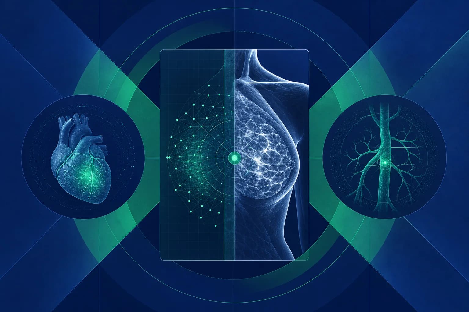
Routine mammography may soon serve a dual purpose in clinical diagnostics by identifying cardiovascular risk factors alongside oncological markers. A study presented at the European Society of Cardiology Congress 2026 demonstrates that a deep learning model can analyze existing mammogram data to flag hypertension, ischemic heart disease, and prior stroke events with significant predictive accuracy.
The research team, led by Dr. Viana Copeland of Tel Aviv University, utilized a dataset comprising 97,000 mammography scans collected from 30,000 women between 2011 and 2025. This large-scale longitudinal data allowed for the training of a neural network designed to identify subtle vascular patterns often overlooked in standard radiological reviews. The model achieved an area under the curve score of 0.79 for hypertension detection, 0.78 for ischemic heart disease, and 0.86 for stroke identification. These metrics indicate a high degree of capability to extract clinically relevant cardiovascular signals from imaging modalities traditionally reserved for breast tissue analysis.
The technical implementation relies on deep learning architectures capable of processing high-resolution medical imagery to detect non-obvious correlations between breast calcification patterns and systemic vascular health. By training the model on a diverse set of patient outcomes, the researchers successfully mapped radiological features to specific cardiovascular conditions. The system effectively functions as a secondary diagnostic layer, providing a non-invasive screening mechanism that integrates into existing clinical workflows without requiring additional patient interaction or radiation exposure.
Data scientists involved in the project emphasized that the model architecture focuses on feature extraction from the entire scan rather than localized regions of interest. This holistic approach ensures that systemic markers, which might otherwise be ignored by human radiologists focused solely on malignancy, are captured by the neural network. The resulting diagnostic output provides a probability score for each of the three cardiovascular conditions, allowing clinicians to prioritize follow-up care for high-risk patients.
The training process involved rigorous cross-validation techniques to ensure the model could generalize across different patient demographics and imaging equipment manufacturers. By utilizing a multi-year dataset, the researchers accounted for variations in image quality and acquisition protocols that typically plague medical imaging datasets. This methodological rigor is essential for moving the technology from a research environment into a validated clinical tool.
Dr. Sonya Babu-Narayan, clinical director at the British Heart Foundation, noted that the integration of such models could address significant disparities in how cardiovascular disease is diagnosed in women. The current diagnostic landscape often suffers from late-stage detection due to persistent clinical biases, where symptoms are frequently misattributed or ignored. By automating the identification of high-risk patients during routine breast screenings, healthcare providers can initiate preventative interventions significantly earlier than current protocols allow.
The research team is currently focused on refining the model to minimize false positive and false negative rates, which remain critical barriers to clinical adoption. Future iterations of the algorithm will likely incorporate broader datasets to improve generalization across different demographic groups and imaging hardware. The researchers are also exploring whether the same deep learning framework can be extended to detect additional heart conditions beyond the initial three identified in the study.
Associate Professor Elena Arbelo, a member of the European Society of Cardiology, emphasized the transition from experimental validation to clinical implementation as the primary challenge for this technology. The potential for mammograms to act as a window into cardiovascular health represents a shift in preventative medicine, where existing diagnostic infrastructure is optimized for multi-pathology detection. This approach highlights the efficacy of applying specialized computer vision models to solve systemic healthcare inefficiencies.
The deployment of these models in a clinical setting would require rigorous regulatory oversight to ensure reliability across diverse hospital environments. Stakeholders are now evaluating the necessary MLOps infrastructure to support real-time inference within radiology departments. Success in this domain depends on the ability to maintain high sensitivity and specificity while ensuring that the automated alerts are integrated into the existing decision-support systems used by cardiologists via standardized API calls.
Future milestones for the project include prospective clinical trials to validate the model’s performance in real-world diagnostic scenarios. If successful, the widespread adoption of this technology could fundamentally alter how cardiovascular risk is screened at scale, particularly for populations that are currently under-diagnosed. The focus remains on establishing the necessary technical and clinical benchmarks to move this research from the conference stage into standard medical practice.